pleural effusion cat ultrasound
This review outlines a practical approach to cases of pleural effusion focusing on early recognition and confirmation of pleural space disease stabilisation of the. Cats with pleural effusion often have rapid shallow breathing and pet owners may notice increased respiratory effort.

Differentiating Pericardial From Pleural Effusion Animal Ultrasound Association
Classification of pleural effusion PE is central to diagnosis.
. In some cases ultrasound may also. 337 including the 5 who had no radiographic evidence of pleural effusion. There are a number of characteristic findings on radiographs that will help your veterinarian identify the presence of pleural effusion.
Abdominal ultrasounds were performed in 70 cats with pleural effusion and revealed concurrent abdominal effusion in 59 of these cats. Pleural effusion can be confirmed with radiography a single DV view if patient permits or thoracic ultrasonography. The lack of specificity is mainly due to the limitations of the imaging modality.
Pleural effusion is typically. Pleural effusion is typically diagnosed by taking radiographs X-rays of the chest. Rishniw M Weidman J Hornof WJ Hydrothorax secondary to a perinephric pseudocyst in a cat.
Transudate protein poorclear watery fluid effusion or. This review outlines a practical approach to cases of pleural effusion focusing on early recognition and confirmation of pleural space disease stabilisation of the patient and logical diagnostic investigation. 694 had obvious pleural effusion and 21183 115 had.
Pleural effusion or pericardial effusion can cause muffled heart sounds. Determining the underlying aetiology is key to appropriate management. All causes of pleural effusion in cats are serious and many carry a.
Vet Radiol Ultrasound 1998. Of these two-thirds 127183. A thoracic chest ultrasound can also help.
The type of pleural fluid withdrawn will enable your veterinarian to diagnose the cause of the pleural effusion. Cats presenting for pleural effusion are often experiencing shortness of breath and decreased oxygen intake placing them into an oxygen cage provides some degree of immediate relief and will allow your cat to calm down enough for a thorough exam and diagnostics. X-ray and ultrasound imaging of the chest cavity are also very helpful in analyzing the causative factors.
Pleural effusion can have a number of different causes including diseases of the heart lungs or other systemic diseases. If the FAST ultrasound does reveal pleural effusion thoracentesis can be carried out. In the latter situations therapeutic intervention must be initiated quickly to prevent respiratory arrest.
The most commonly diagnosed cause of pleural effusion in cats is chylothorax. Found with right congestive heart failure obstruction to lymphatic drainage by tissue adhesions in pleural space lung lobe torsion neoplasms and abdominal contents herniating. Given that most effusions are detected by x-ray which generally cannot distinguish between fluid types the fluid in.
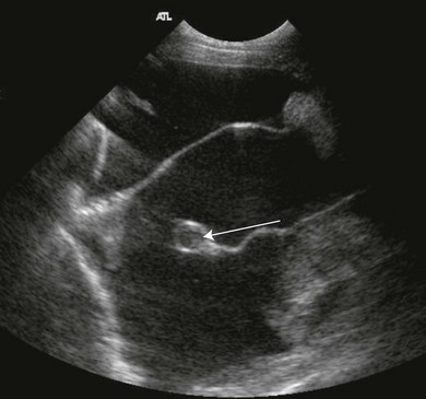
Approximately half of the cats 183380. There is some fluid in the pericardium but this is normal and only seen during systole. Determining the underlying aetiology is.
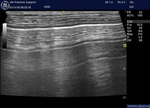
Pleural effusion was confirmed in all of the cats who had thoracic ultrasonography 128380. The L marks where an air filled lung lobe should be. Retrospective analysis of pleural effusion in cats CD and neoplasia were the most common causes for feline pleural effusion.
Accumulation of fluid in the pleural space. Cats may develop open-mouthed breathing in an effort to increase air flow. Major Differential Diagnoses for Pleural Effusion in the Cat.
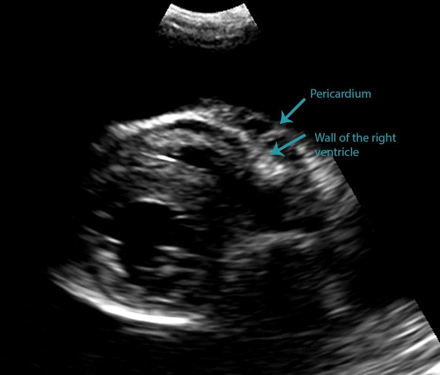
In the moving clip however you can actually see the separation of the right ventricular free wall from the pericardium in a cat. In many cases thoracocentesis is used to remove accumulated fluid. CD and neoplasia were the most common causes for feline pleural effusion.
Diverse disease processes result in sufficient fluid accumulation within the pleural space to cause respiratory compromise. In human medicine PEs are divided into only two categories. Four standard effusion types recognized in addition to blood.
Testing that can help with diagnosis includes blood work urinalysis x-ray ultrasound and evaluation of fluid collected from the chest with a needle. Cats with pleural effusion often have severe respiratory compromise at presentation. The aim of this study was to evaluate in 20 cats presented with PE paired samples of serum and pleural.
Diverse disease processes result in sufficient fluid accumulation within the pleural space to cause respiratory compromise. Pleural effusion in cats with pyothorax in. A sample of pleural fluid obtained by piercing the cats chest cavity with a needle will be sent to the laboratory for analysis.
Some affected cats may also cough. Traditional veterinary classification has distinguished between transudates modified transudates and exudates. 482 had echocardiographic studies.
Cats presenting with pleural effusion are nearly always in respiratory distress ranging from an increased respiratory rate and effort to open mouth breathing. Several medical conditions are responsible for pleural effusion PLEFF with volume overload congestive heart failure and pleuropulmonary infection representing the most common causes in the intensive care unit ICU Fluid accumulation can take place due to an imbalance of hydrostatic and oncotic pressure across the lung capillaries. The therapeutic intervention also provides your first diagnostic test.
In this cat below it is difficult at first glance to be able to state whether this effusion is pleural or pericardial. Age liver enzymes as well as cell count protein and glucose levels in the effusion can aid in the investigation of underlying aetiologies. This can be caused by thoracic lymphangiectasia swollen lymph vessels that leak chyle into the pleural space congestive heart failure obstruction of the cranial vena cava the major vein that returns blood to the heart from the front of the body cancer fungal infection feline heartworm.
Pleural effusion is commonly used as a catch-all term to describe any abnormal accumulation of fluid in the pleural cavity. This procedure removes excess fluid from the pleural space using a needle which not only relieves pressure on your cats lungs but also provides your. An x-ray is helpful in diagnosing pleural effusion because the abnormal fluid can be seen in the chest cavity.
In some cases ultrasound may also be. Collection of pleural effusion was performed by blind or ultrasound-guided thoracentesis. Examination of the effusion included determination of specific gravity using a refractometer Atago Company as well as measurement of the total cell count with the Cell-Dyn 3500 System Abott Laboratories.
Once fluid is identified in the chest cavity the cause must be determined. Abdominal abnormalities identified on ultrasound included abdominal masses lymphadenopathy hepatic venous congestion hepatomegaly splenomegaly renal enlargement small intestinal wall thickening steatitis and pancreatitis.

Pleural Effusion Chest X Rays Radiology Pediatric Medicine Icu Nursing

Pleural Effusion Radiology Reference Article Radiopaedia Org
Lung Ultrasound Flooding In Fulminant Pulmonary Oedema In Cats And A Comparison With Pneumonia Vet Practice Support
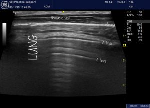
Lung Ultrasound Contusions Interstitial Syndrome A B C E I And Z Lines Explored Vet Practice Support

Cardioechography Ultrasound Physics Ultrasound School Sonography Student

Midscapular Thoracentesis Ultrasound Training Model Ultrasound Training Ultrasound Emergency Medicine

Sonography Assessment Overview Of Afast And Tfast Today S Veterinary Practice

Sonography Assessment Overview Of Afast And Tfast Today S Veterinary Practice

How To Ultrasound Detection Of Pleural Fluid Case Study Video Youtube
Differentiating Pericardial From Pleural Effusion Animal Ultrasound Association

Different Types Of Pleural Effusion On Ultrasound Scan A Exudate B Download Scientific Diagram

Veterinary Echocardiography Newsletter 1 Effusions Animal Ultrasound Association

Midscapular Thoracentesis Ultrasound Training Model Ultrasound Training Ultrasound Emergency Medicine

Pin By Sal Thompson On Radiography Medical Ultrasound Vision Eye Radiography
Pleura Veterian Key

Sonography Assessment Overview Of Afast And Tfast Today S Veterinary Practice

Differentiating Pericardial From Pleural Effusion Animal Ultrasound Association

Gangrenous Cholecystitis Radiology Case Radiopaedia Org Medical Ultrasound Ultrasound Sonography Radiology

Animal Jobs Near Me 2022 Veterinary Radiology Radiology Online Textbook